The knee has four main ligamentous structures that act to stabilise the joint and limit excess movement. One of these ligaments is known as the Lateral Collateral Ligament, or the LCL for short. The LCL runs over the outside of the knee and attaches the thigh bone (femur) to the bone found on the outside of the shin (the fibula). Due to its attachment points on the outside of the knee, the role of the lateral collateral ligament is to prevent the knee from collapsing outward (this is known as lateral, or varus collapse). Due to the demands of their respective sports, LCL injuries are most common in tennis players and gymnasts.
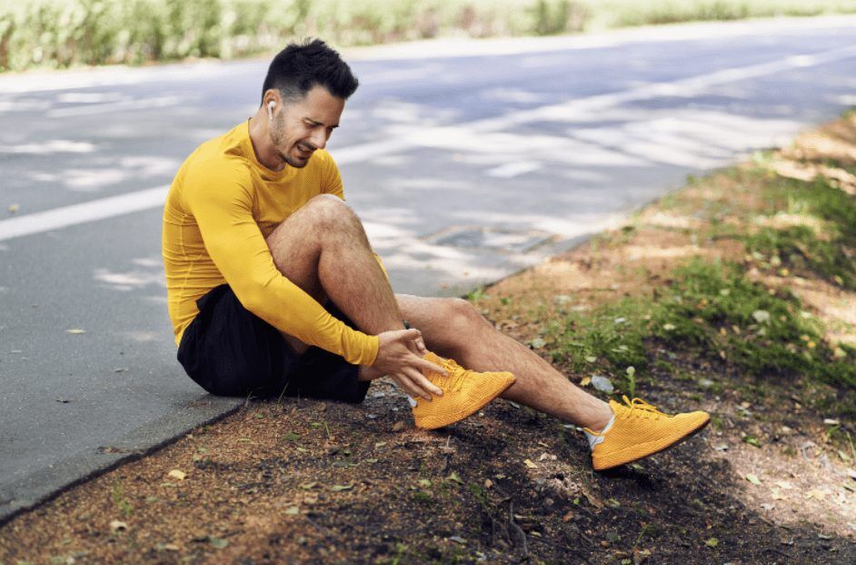
How do I injure my LCL?
It is quite unusual to see an isolated lateral collateral ligament injury (these represent less than 2% of total knee injuries). A direct blow to the inside of the knee, forcing it to bend outwards is the most common mechanism for this type of injury. In some scenarios, hyperextension of the knee joint has also been known to harm the LCL. Because of the force required to damage a lateral collateral ligament, it is common to see anterior cruciate ligament, posterior cruciate ligament and other associated injuries simultaneously.
What are the symptoms of an LCL injury?
An injury to your Lateral Collateral Ligament will usually result from a varus-type injury, as mentioned above. Pain, swelling, and tenderness over the outside of the knee, where the thigh and shin bones meet is also common. Due to the swelling & inflammation, your knee range of motion may be reduced, and you may struggle to weight bear on the affected leg. Finally, you may report to your physiotherapist that you felt a “pop” over the outside of the knee at the time of injury.
How is an LCL injury diagnosed?
The first step in diagnosing any knee injury is an assessment with a health professional, such as a physiotherapist or emergency physician. They will perform a number of physical tests to assess the integrity and stability of the affected ligament. Should your physiotherapist find it necessary, they can order an x-ray of the knee to assess for potentially associated fractures. The gold standard of imaging for ligamentous injuries is an MRI, which can be requested to accurately assess the severity and location of your injury.
How is an LCL injury treated?
The treatment of a Lateral Collateral Ligament injury is dependent on the grade of injury sustained. LCL injuries are graded from I to III, with Grade I injuries involving stretching of the ligamentous fibers. A partial tear of the LCL is considered a grade II injury, while a grade III injury describes a complete rupture of the ligament.
Grade I & II Lateral Collateral Ligament injuries are managed conservatively. This can involve bracing the knee in a particular range of motion for a period of time, as well as specific exercises prescribed by your physiotherapist. It can take at least 6-8 weeks to return to sport after an injury of this nature. A grade III injury or complete rupture of the Lateral Collateral Ligament is generally managed surgically, with a graft taken from one of the hamstring tendons. The best results are seen when the surgery is performed quite quickly (within 2 weeks) of the injury being sustained.