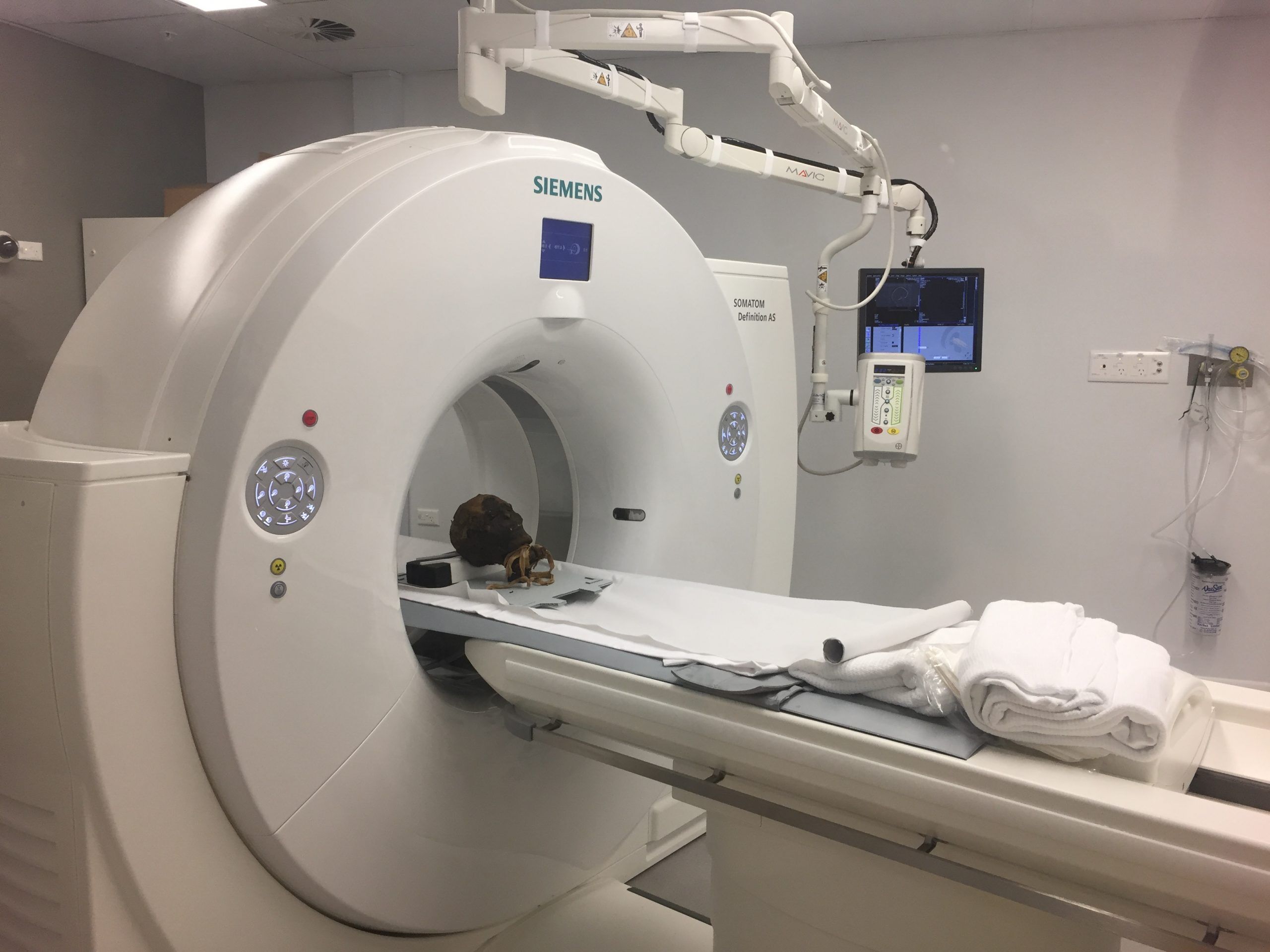
Mummy Observed via CT imaging
An Egyptian mummy artefact from the Nicholson Museum was scanned using a Siemens SOMATOM AS+.
The artefact was scanned using both a high resolution single source technique as well as dual energy to reveal as much information as possible for thorough investigation using forensic imaging techniques. Egyptologists Dr Janet Davey and Dr Karin Sowada will both work through the data to determine as much as possible about the life of this man who died more than 2000 thousand years ago in the Ptolemaic period.
CT imaging at such a high resolution has become an invaluable tool for the non-destructive investigation into ancient artefacts and helps researchers to better understand our history as a race. By working closely with Siemens, University research staff also had access to Cinematic Rendering, a unique approach to photo-realistic image rendering of image data such as CT and MRI. This method generates much more life-like images from which to draw and preserve information for posterity.

